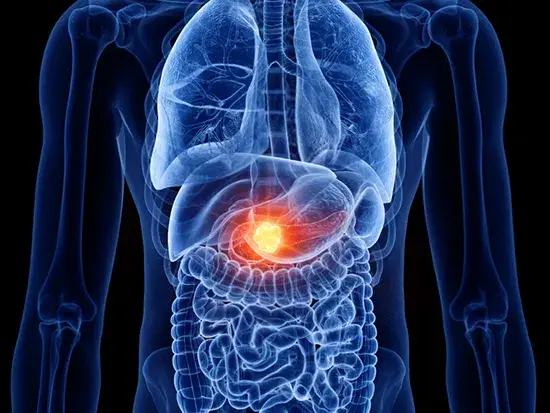
Early Warning Signs of Cancer Growth and the Surprising Role of Blood Type in Your Cancer Risk
Introduction
Cancer remains one of the leading causes of mortality worldwide, in part because many forms progress silently until they reach advanced stages. Detecting malignancies early can dramatically improve treatment options and outcomes—but that requires knowing which subtle changes in your body merit further investigation. In this comprehensive guide, we will:
-
Explore five early warning signs that can indicate cancerous growth.
-
Delve into the science behind each symptom, including which cancers are most often associated with them.
-
Examine the intriguing link between ABO blood types and cancer risk, highlighting why individuals with Type O blood may enjoy a relative protective advantage.
-
Offer practical advice on when and how to seek medical evaluation if you experience any of these warning signs.
By the end of this article, you will have a clear, actionable understanding of how to monitor your health for early red flags and empower yourself with knowledge that could accelerate diagnosis, facilitate timely treatment, and ultimately save lives.
1. Unexplained Weight Loss: The Body’s Early Distress Signal
1.1 Recognizing the Pattern
It is normal to experience minor fluctuations in weight—perhaps a few pounds gained after a vacation or lost during a particularly stressful week. However, when you lose 10 pounds (approximately 4.5 kilograms) or more over a 6– to 12–month period without intentional changes to your diet or activity level, this warrants a closer look.
1.2 Why Cancer Causes Rapid Weight Loss
-
Metabolic Disruption: Cancer cells proliferate rapidly, consuming large quantities of glucose and amino acids. This heightened energy demand can shift the body into a hypermetabolic state, burning more calories at rest.
-
Cachexia: In advanced malignancies, a syndrome known as cachexia may develop, characterized by muscle wasting and fat loss that cannot be fully reversed with nutrition alone.
-
Digestive Interference: Tumors in the gastrointestinal tract—such as those in the stomach, pancreas, or liver—can obstruct food passage, impair nutrient absorption, and reduce appetite.
1.3 Types of Cancer Commonly Associated
-
Gastric (Stomach) Cancer: Tumors in the stomach lining can cause early satiety and nausea, leading to unintended weight loss.
-
Pancreatic Cancer: Often dubbed “the silent killer,” pancreatic tumors frequently manifest with weight loss as one of the first signs, due to both obstructive symptoms and systemic metabolic effects.
-
Liver Cancer: Primary or metastatic lesions in the liver can impair detoxification and nutrient processing, resulting in appetite loss and weight decline.
-
Esophageal Cancer: Tumors in the esophagus can create swallowing difficulties (dysphagia), prompting patients to eat less and lose weight.
-
Hematologic Malignancies (Leukemia, Lymphoma): These cancers of the blood and lymph nodes can cause systemic inflammation and metabolic disturbances that drive rapid weight loss.
1.4 What to Do
-
Track Your Trends: Record your weight weekly under similar conditions (e.g., same time of day, similar clothing).
-
Review Lifestyle Factors: Confirm that no recent changes in diet, exercise, stress levels, or medications could explain the loss.
-
Consult a Physician: If unintentional weight loss exceeds 5% of your body weight over six months, seek medical evaluation. Your doctor may order blood panels, imaging studies (e.g., abdominal ultrasound or CT scan), or endoscopic procedures to identify potential causes.
2. Persistent Fatigue and Weakness: More Than Just Tiredness
2.1 Distinguishing “Normal” Fatigue from Concerning Exhaustion
Everyone feels tired after a long day or a sleepless night. Cancer–related fatigue, however, is:
-
Constant: Present upon waking and persisting throughout the day.
-
Disproportionate: Far more severe than what you would expect from your level of activity or rest.
-
Non-Relieving: Does not improve substantially with sleep, nutrition, or short breaks.
2.2 Mechanisms Underlying Cancer–Related Fatigue
-
Anemia: Many cancers—particularly leukemia and lymphoma—interfere with red blood cell production, reducing the blood’s oxygen–carrying capacity and leading to profound tiredness.
-
Cytokine Release: Tumors can stimulate chronic inflammation, releasing cytokines that disrupt sleep patterns and energy metabolism.
-
Metabolic Dysregulation: As with weight loss, hypermetabolism driven by tumor activity can exhaust the body’s fuel reserves.
-
Psychological Toll: Anxiety about health, pain, and treatment side effects can further compound physical fatigue.
2.3 Associated Cancer Types
-
Hematologic Cancers: Leukemia and lymphoma frequently present with severe fatigue due to bone marrow infiltration and blood panel abnormalities.
-
Gastrointestinal Tumors: Colon and stomach cancers can cause chronic, low–grade bleeding—leading to iron–deficiency anemia and resultant weakness.
-
Hepatic (Liver) Malignancies: Liver tumors compromise detoxification pathways and nutrient storage, contributing to systemic fatigue.
2.4 Recommended Actions
-
Keep an Exhaustion Log: Note the time, duration, and severity of fatigue episodes, as well as any alleviating or aggravating factors.
-
Investigate for Anemia: A complete blood count (CBC) can reveal low hemoglobin or hematocrit levels indicating anemia.
-
Screen for Inflammatory Markers: Elevated C–reactive protein (CRP) or erythrocyte sedimentation rate (ESR) may point to underlying inflammatory or malignant processes.
-
Seek Early Medical Advice: If fatigue interferes with daily activities—or if it is accompanied by other warning signs such as night sweats, fevers, or weight loss—schedule a physician visit for a comprehensive workup.
3. Skin Changes: The Body’s Visible Distress Signals
3.1 Beyond Sunburn: Recognizing Concerning Dermatological Signs
While many skin changes are harmless—aging spots, eczema, or simple bruises—some warrant professional evaluation when they:
-
Evolve Rapidly: Change in size, shape, color, or texture over weeks to months.
-
Refuse to Heal: Lesions or ulcers that persist or worsen despite standard care.
-
Appear in Unusual Patterns: Discrete marks in non–sun–exposed areas or accompanied by systemic symptoms.
3.2 Common Skin Warning Signs and Their Implications
| Skin Sign | Possible Underlying Cancer |
|---|---|
| Asymmetrical, irregularly bordered, multicolored mole | Melanoma |
| Non–healing sores or ulcers | Squamous cell carcinoma, basal cell carcinoma |
| Jaundice (yellowing of skin and whites of eyes) | Pancreatic cancer, liver cancer, bile duct obstruction |
| Sudden appearance of multiple dark spots or itching | Paraneoplastic syndromes (e.g., related to internal malignancies) |
3.3 The Science Behind Dermatological Indicators
-
Melanoma Development: Mutations in melanocytes can drive uncontrolled proliferation, manifesting as rapidly changing moles.
-
Chronic Ulceration: Loss of normal skin architecture in precancerous lesions can lead to ulcer formation when repair mechanisms fail.
-
Jaundice Mechanism: Tumors in the hepatobiliary tract can block bile flow, causing bilirubin accumulation and yellow discoloration.
-
Paraneoplastic Effects: Some internal cancers release hormones or cytokines that trigger widespread skin manifestations (e.g., acanthosis nigricans may signal gastric adenocarcinoma).
3.4 Best Practices for Monitoring Skin Health
-
Monthly Self–Exams: Use the ABCDE rule for moles—Asymmetry, Border irregularity, Color variation, Diameter >6mm, Evolving characteristics.
-
Annual Dermatologist Visits: Especially if you have a history of sunburn, tanning bed use, or a family history of skin cancer.
-
Prompt Biopsy: Any suspicious lesion that bleeds easily, itches persistently, or fails to heal within 4–6 weeks should be biopsied.
4. Persistent Pain: When Discomfort Signals Something Deeper
4.1 Distinguishing Chronic Pain from Cancer–Related Pain
While chronic pain can arise from arthritis, nerve damage, or overuse injuries, cancer–related pain often:
-
Persists Without Obvious Cause: No identifiable injury, infection, or degenerative disease.
-
Escalates Over Time: Gradually intensifies in severity and frequency.
-
Resists Standard Treatments: Does not respond adequately to over–the–counter analgesics or rest.
4.2 How Tumors Generate Pain
-
Direct Invasion: Growth into bone (bone cancer), visceral organs, or nerve sheaths can produce localized, deep–seated pain.
-
Mass Effect: Pressure from a growing mass can compress nearby structures—e.g., colorectal tumors causing abdominal cramping.
-
Inflammatory Mediators: Cancer cells release prostaglandins and cytokines, sensitizing pain receptors.
4.3 Cancers Frequently Presenting with Pain
| Pain Location | Potential Cancer Types |
|---|---|
| Deep bone pain | Primary bone cancers (osteosarcoma, Ewing sarcoma), metastatic lesions |
| Persistent headaches | Brain tumors (gliomas, meningiomas), metastatic disease |
| Pelvic or abdominal pain | Ovarian, colorectal, or pancreatic cancers |
| Chest discomfort | Lung cancer, esophageal tumors |
4.4 When to Seek Evaluation
-
Duration Threshold: Pain lasting more than 4–6 weeks without improvement.
-
Red–Flag Features: Night–time pain that awakens you, unintentional weight loss alongside pain, neurological deficits (numbness, weakness), or gastrointestinal bleeding.
-
Recommended Workup: Imaging studies (X–ray, MRI, CT), bone scans, endoscopy, or referral to an oncologist depending on the pain’s location and character.
5. Unusual Lumps or Swellings: Palpable Clues to Underlying Growths
5.1 The Importance of Early Detection
Identifying a new lump or swelling early gives you the best chance of curative treatment. While not every lump is malignant—cysts, lipomas, or reactive lymph nodes are common—certain features raise suspicion:
-
Firmness: Hard or rock–like consistency.
-
Painlessness: Some benign lumps are tender; many cancerous nodules are not.
-
Progressive Growth: An increase in size over weeks or months.
5.2 Common Sites and Associated Malignancies
| Location | Possible Cancer |
|---|---|
| Breast or armpit | Breast carcinoma |
| Testicle | Testicular germ cell tumors |
| Neck (thyroid region) | Thyroid carcinoma, lymphoma |
| Lymph node regions (groin, neck, armpit) | Lymphoma, metastatic carcinomas |
5.3 Pathophysiology of Malignant Nodules
-
Uncontrolled Cell Division: Cancer cells bypass normal growth checkpoints, creating masses of abnormal tissue.
-
Angiogenesis: Tumors recruit new blood vessels, which can cause swelling and visible veins overlying the mass.
-
Local Invasion: Malignant cells can infiltrate surrounding structures, making the lump fixed rather than freely mobile.
5.4 Action Steps for Any New Lump
-
Self–Examination: Monthly breast or testicular self–checks, noting any changes in texture or feel.
-
Clinical Assessment: A physician may perform an ultrasound–guided fine needle aspiration (FNA) or core biopsy to obtain tissue samples.
-
Imaging: Mammogram, scrotal ultrasound, CT, or PET scans may be indicated to evaluate extent and characteristics.
6. The Surprising Link Between Blood Type and Cancer Risk
6.1 Overview of the ABO Blood Group System
The ABO system classifies blood into four primary groups—A, B, AB, and O—based on the presence or absence of specific antigens on red blood cells. Each group can further be Rh–positive or Rh–negative, yielding eight possible combinations (e.g., O–positive, A–negative).
6.2 Historical Investigations
Researchers have long explored whether blood group antigens influence susceptibility to certain diseases. Hypotheses include:
-
Cellular Adhesion: Blood group antigens may affect how cells—including malignant ones—adhere to each other and to blood vessel walls.
-
Immune Surveillance: Variations in antigen expression could modulate immune recognition of cancer cells.
-
Microbiome Interactions: Blood group–related differences in gut flora might impact colorectal or gastric cancer risk.
7. Why Type O Blood May Confer a Protective Advantage
7.1 Key Study Findings
A landmark 2015 meta–analysis examined thousands of patients worldwide and found that individuals with Type O blood exhibited:
-
Reduced Risk of Gastric Cancer: Up to a 20% lower incidence compared to non–O groups.
-
Lower Rates of Pancreatic Cancer: Approximately 13% decreased risk relative to Types A, B, and AB.
-
Decreased Incidence of Colorectal Cancer: A modest but statistically significant protective effect.
7.2 Proposed Mechanisms
-
Reduced Clotting Propensity: Non–O blood types have higher levels of von Willebrand factor and clotting factors, which may contribute to microthrombi formation that fosters a pro–tumorigenic environment.
-
Inflammatory Mediators: Type O individuals often have lower baseline levels of certain inflammatory markers, potentially curbing chronic inflammation—a known driver of carcinogenesis.
-
H. pylori Interaction: The bacterium Helicobacter pylori, linked to gastric cancer, binds more avidly to gastric mucosa in non–O individuals, elevating infection severity.
7.3 Clinical Implications
-
Risk Stratification: While blood type alone is not sufficient to dictate screening frequency, it may complement other risk factors (family history, lifestyle habits) in personalized prevention plans.
-
Preventive Counseling: Type O individuals should still adhere to standard cancer screening guidelines but may be reassured of their slightly lower statistical risk for certain tumors.
8. Limitations and Caveats: Blood Type Is Not Destiny
8.1 Multifactorial Nature of Cancer
Cancer risk arises from the interplay of genetics, environment, lifestyle, infections, and random cellular mutations. Blood type constitutes only one minor dimension of a vastly complex risk profile.
8.2 Conflicting Evidence
Subsequent studies have produced mixed results:
-
Some found no significant association between ABO group and breast or lung cancer.
-
Others reported population–specific differences, suggesting that genetic background and regional factors modulate any blood–type effect.
8.3 Avoiding False Reassurance
Whether your blood type is O or AB, the cornerstones of cancer prevention remain:
-
Healthy Diet: Emphasize fruits, vegetables, whole grains, and lean proteins.
-
Regular Exercise: Aim for at least 150 minutes of moderate activity per week.
-
Tobacco and Alcohol Moderation: Eliminate smoking and limit alcohol intake.
-
Vaccinations: HPV and hepatitis B vaccines guard against virus–related cancers.
-
Standard Screenings: Follow age– and risk–appropriate guidelines for mammograms, colonoscopies, Pap tests, and low-dose CT for smokers.
9. The Power of Early Detection and Proactive Management
9.1 Screening Saves Lives
-
Breast Cancer: Mammography detects non–palpable tumors before they spread.
-
Colorectal Cancer: Colonoscopy removes precancerous polyps, preventing progression.
-
Cervical Cancer: Pap smears and HPV testing intercept dysplasia early.
-
Lung Cancer: Low–dose CT scans in high–risk smokers reduce mortality.
9.2 Symptom–Driven Evaluation
Even with rigorous screening, symptoms remain critical triggers for further investigation. Should you experience any of the five warning signs described above—unexplained weight loss, persistent fatigue, skin changes, chronic pain, or new lumps—do not delay consulting your healthcare provider.
9.3 Building a Partnership with Your Physician
-
Maintain Records: Share explicit details about symptom onset, duration, and progression.
-
Ask Questions: Inquire about recommended diagnostic tests, potential side effects, and next steps.
-
Follow Through: Attend follow–up appointments and adhere to specialist referrals promptly.
10. Practical Guide: When and How to Seek Medical Attention
| Symptom | Action Steps |
|---|---|
| Unexplained weight loss >5% body weight | Schedule a primary care visit; obtain a CBC, metabolic panel, and possibly imaging studies. |
| Constant, non–relieving fatigue | Rule out anemia (CBC), thyroid dysfunction (TSH), and inflammatory conditions (CRP, ESR). |
| Concerning skin changes | Photograph lesions monthly; refer to a dermatologist for dermoscopy and possible biopsy. |
| Persistent pain without clear cause | Start with localized imaging (X–ray, ultrasound); escalate to MRI or CT if pain persists beyond 4 weeks. |
| New or growing lump/swelling | Arrange ultrasound or CT scan; consider needle biopsy if imaging suggests malignancy. |
11. Conclusion
Cancer’s stealthy progression underscores the importance of vigilance. By familiarizing yourself with five key early warning signs—unexplained weight loss, chronic fatigue, troubling skin changes, persistent pain, and unusual lumps—you equip yourself to recognize when “normal” bodily fluctuations cross into potentially dangerous territory. Additionally, understanding that your ABO blood type, particularly Type O, may slightly influence your statistical cancer risk can inform—but must not dictate—your preventive health strategy.
Ultimately, early detection remains the most powerful tool in the fight against cancer. Combine regular screenings, a healthy lifestyle, and prompt medical attention for concerning symptoms to maximize your chances of favorable outcomes. If you experience any of the signs described in this guide, reach out to your healthcare provider without delay—because catching cancer early can make all the difference.
References & Further Reading
-
National Cancer Institute, “Understanding Cancer,” cancer.gov
-
World Health Organization, “Cancer Fact Sheet,” who.int
-
American Cancer Society, “Cancer Prevention & Early Detection,” cancer.org
-
Meta-Analysis on ABO Blood Types and Cancer Risk (2015)
Disclaimer: This article is for informational purposes only and does not substitute professional medical advice. Always consult a qualified healthcare provider for diagnosis and treatment.

Lila Hart is a dedicated Digital Archivist and Research Specialist with a keen eye for preserving and curating meaningful content. At TheArchivists, she specializes in organizing and managing digital archives, ensuring that valuable stories and historical moments are accessible for generations to come.
Lila earned her degree in History and Archival Studies from the University of Edinburgh, where she cultivated her passion for documenting the past and preserving cultural heritage. Her expertise lies in combining traditional archival techniques with modern digital tools, allowing her to create comprehensive and engaging collections that resonate with audiences worldwide.
At TheArchivists, Lila is known for her meticulous attention to detail and her ability to uncover hidden gems within extensive archives. Her work is praised for its depth, authenticity, and contribution to the preservation of knowledge in the digital age.
Driven by a commitment to preserving stories that matter, Lila is passionate about exploring the intersection of history and technology. Her goal is to ensure that every piece of content she handles reflects the richness of human experiences and remains a source of inspiration for years to come.
