Your skin tells a story—of years spent in the sun, genetic inheritance, and the natural aging process. Sometimes, this story manifests as changes that might seem insignificant at first glance but deserve closer attention. Rough patches, scaly areas, or unusual growths can be more than cosmetic concerns; they can be windows into your skin’s health and indicators of what care it needs. Understanding these changes and knowing when to take action could make the difference between a simple treatment and a more serious health concern.
What Is Keratosis? Understanding the Foundation
Keratosis represents a family of skin conditions characterized by the overproduction of keratin—a crucial protein that forms the structural foundation of your skin, hair, and nails. Think of keratin as your skin’s natural armor, providing protection and structure to the outermost layers of your body’s largest organ.
When this protein production goes into overdrive, it creates a buildup that manifests as rough, scaly, or thickened patches on the skin’s surface. These formations can vary dramatically in appearance, location, and significance, ranging from purely cosmetic concerns to potentially serious precancerous conditions that require immediate medical attention.
The overproduction of keratin doesn’t happen in isolation—it’s typically the result of various triggers including prolonged sun exposure, genetic predisposition, hormonal changes, or underlying skin conditions. Understanding these underlying mechanisms helps explain why keratosis affects different people in different ways and why treatment approaches must be tailored to individual circumstances.
What makes keratosis particularly important to understand is its spectrum of severity. While many forms are entirely benign and pose no health risks beyond cosmetic concerns, others serve as early warning signs of potentially dangerous skin changes. This variability underscores the importance of proper identification and professional evaluation.
The Many Faces of Keratosis: Types and Characteristics
Seborrheic Keratosis: The “Barnacles of Aging”
Seborrheic keratosis represents one of the most common skin growths encountered in dermatology practices worldwide. These benign lesions have earned the colloquial nickname “barnacles of aging” due to their tendency to appear and multiply as people get older, much like barnacles accumulating on a ship’s hull over time.
These growths present a distinctive appearance that experienced dermatologists can often identify at a glance. They typically manifest as brown, black, or tan spots with a characteristic waxy, “stuck-on” appearance that looks as though someone applied a piece of candle wax to the skin. The surface often appears warty or rough, with a texture that feels different from the surrounding normal skin.
The size of seborrheic keratoses can vary significantly, from tiny spots barely noticeable to large patches several centimeters in diameter. They can appear virtually anywhere on the body, though they’re particularly common on the chest, back, shoulders, and face. Unlike many other skin conditions, seborrheic keratoses tend to multiply over time, with some individuals developing dozens or even hundreds of these growths as they age.
What’s particularly reassuring about seborrheic keratoses is their benign nature. They never transform into cancer and pose no health risks beyond occasional irritation if they’re located in areas where clothing rubs against them or if they’re accidentally scratched or injured. This benign nature doesn’t mean they should be ignored entirely, however, as they can sometimes be confused with more serious skin conditions, including melanoma.
The exact cause of seborrheic keratoses remains somewhat mysterious, though researchers have identified several contributing factors. Genetics clearly plays a significant role, as these growths tend to run in families. Some people inherit a predisposition to develop numerous seborrheic keratoses, while others may develop only a few throughout their lifetime.
Age is the most consistent risk factor, with seborrheic keratoses becoming increasingly common after age 50. However, they can appear earlier in life, particularly in individuals with a strong family history. Hormonal changes, particularly those associated with pregnancy, may also trigger their development or cause existing ones to grow more rapidly.
Actinic Keratosis: The Sun’s Unwelcome Gift
Actinic keratosis, also known as solar keratosis, represents a completely different category of skin change—one that demands serious attention and prompt treatment. Unlike the benign seborrheic keratosis, actinic keratosis is considered a precancerous condition that can potentially progress to squamous cell carcinoma, a form of skin cancer.
These lesions appear as rough, dry, or scaly patches that feel like sandpaper to the touch. They typically range in color from pink to red to brown, and their texture is their most distinguishing feature. Many people describe them as patches of skin that feel perpetually dry or rough, no matter how much moisturizer is applied.
The distribution of actinic keratoses tells their origin story clearly—they appear almost exclusively on areas of the body that receive the most sun exposure over a lifetime. The face, ears, scalp (particularly in men with male-pattern baldness), hands, and forearms are the most common locations. This distribution pattern directly reflects decades of cumulative ultraviolet (UV) radiation exposure.
What makes actinic keratoses particularly concerning is their potential for malignant transformation. While not every actinic keratosis will progress to cancer, studies suggest that approximately 10-15% may eventually develop into squamous cell carcinoma if left untreated. This risk varies based on individual factors including immune system status, continued sun exposure, and genetic susceptibility.
The development of actinic keratoses follows a predictable pattern related to sun exposure history. People who spent significant time outdoors for work or recreation, particularly during their younger years, are at highest risk. Fair-skinned individuals, those with light-colored eyes and hair, and people who burn easily rather than tan are particularly susceptible.
Interestingly, actinic keratoses often appear in clusters or fields, reflecting areas of widespread sun damage. Dermatologists refer to this phenomenon as “field cancerization,” recognizing that the visible lesions may represent only a portion of the sun-damaged tissue in a given area.
Keratosis Pilaris: The “Chicken Skin” Condition
Keratosis pilaris presents an entirely different picture from its keratosis cousins, both in appearance and significance. This common condition affects up to 40% of adults and an even higher percentage of children and teenagers, making it one of the most prevalent skin conditions worldwide.
The hallmark of keratosis pilaris is the appearance of small, rough bumps that give the skin a texture reminiscent of chicken skin—hence its common nickname. These bumps are typically white or red and are most commonly found on the arms, thighs, cheeks, and buttocks. The condition occurs when keratin plugs hair follicles, creating small, raised bumps around each hair.
Unlike other forms of keratosis, keratosis pilaris is primarily a cosmetic concern rather than a health issue. It poses no risk of cancer or other serious complications, though it can cause significant psychological distress, particularly in teenagers and young adults who may feel self-conscious about their appearance.
The condition has a strong genetic component, with many affected individuals having family members with the same condition. It’s also closely associated with other skin conditions, particularly atopic dermatitis (eczema) and ichthyosis. People with these conditions are more likely to develop keratosis pilaris, suggesting shared underlying mechanisms.
Environmental factors also play a role in keratosis pilaris severity. Dry weather, low humidity, and harsh soaps can worsen the condition, while moisture and gentle skin care can help improve it. The condition often fluctuates seasonally, becoming worse in winter when humidity is low and improving in summer when skin tends to be more moist.
One encouraging aspect of keratosis pilaris is its tendency to improve with age. Many children and teenagers who are significantly affected find that their symptoms diminish in adulthood, though some degree of the condition may persist throughout life.
Understanding the Underlying Causes
The Role of Sun Exposure
Ultraviolet radiation from the sun represents the single most significant environmental factor in the development of certain types of keratosis, particularly actinic keratosis. Understanding how UV exposure leads to skin changes helps explain why prevention is so crucial and why certain populations are at higher risk.
UV radiation damages skin in multiple ways, but its effect on DNA in skin cells is particularly relevant to keratosis development. When UV rays penetrate the skin, they can cause direct DNA damage in keratinocytes—the cells that produce keratin. This damage can disrupt normal cell growth and division, leading to the overproduction of keratin that characterizes keratosis.
The relationship between sun exposure and actinic keratosis isn’t simply about total exposure—it’s also about cumulative exposure over time. Decades of moderate sun exposure can be just as damaging as shorter periods of intense exposure. This explains why actinic keratoses typically appear later in life, representing the cumulative effect of years or decades of UV exposure.
Different types of UV radiation have different effects on the skin. UVB radiation primarily affects the outer layers of skin and is responsible for sunburn, while UVA radiation penetrates deeper and contributes to premature aging and skin damage. Both types contribute to keratosis development, which is why broad-spectrum sun protection is essential.
The intensity of UV exposure varies based on multiple factors including geographic location, altitude, time of day, season, and weather conditions. People living closer to the equator, at higher altitudes, or in areas with intense sun reflection (like snow or water) face higher UV exposure risks.
Genetic Predisposition
Genetics plays a crucial role in determining who develops keratosis and what types they’re likely to encounter. This genetic influence operates through multiple mechanisms, from determining skin type and color to influencing how efficiently DNA repair mechanisms function.
Skin color, determined by melanin production, significantly affects keratosis risk. People with fair skin, light hair, and light eyes have less natural protection against UV radiation and are more susceptible to developing actinic keratosis. This genetic predisposition to sun sensitivity can be traced to evolutionary adaptations to different geographic regions and sun exposure levels.
For seborrheic keratosis, genetic factors appear to influence both the tendency to develop these growths and their eventual number and distribution. Some families show clear patterns of inheritance, with multiple generations affected by numerous seborrheic keratoses. While the specific genes involved are still being studied, the familial clustering is unmistakable.
Keratosis pilaris shows perhaps the strongest genetic component of all keratosis types. It’s inherited in an autosomal dominant pattern, meaning that if one parent has the condition, there’s a significant chance their children will develop it as well. The condition is also linked to mutations in genes involved in skin barrier function and hair follicle development.
Understanding genetic predisposition doesn’t mean that developing keratosis is inevitable—environmental factors and personal choices still play crucial roles. However, knowing your genetic risk can help guide prevention strategies and screening recommendations.
Environmental and Lifestyle Factors
Beyond sun exposure and genetics, numerous environmental and lifestyle factors influence keratosis development and severity. These factors often interact with genetic predisposition in complex ways, either amplifying or mitigating risk.
Dry skin conditions can worsen all types of keratosis, particularly keratosis pilaris. Low humidity environments, harsh soaps, hot water exposure, and inadequate moisturizing can all contribute to skin dryness and worsen keratosis symptoms. This explains why many people notice their keratosis becomes more prominent during winter months or when living in arid climates.
Occupational exposures can significantly impact keratosis risk. People who work outdoors—construction workers, farmers, lifeguards, and others—face elevated UV exposure that increases actinic keratosis risk. Similarly, exposure to certain chemicals or irritants in workplace settings may contribute to skin changes.
Hormonal changes can influence keratosis development and progression. Pregnancy, in particular, seems to trigger or worsen seborrheic keratosis in some women. This hormonal influence may explain why certain types of keratosis show gender-related differences in prevalence and severity.
Immune system status also plays a role, particularly for actinic keratosis. People with compromised immune systems—whether due to medications, medical conditions, or age-related immune decline—may develop actinic keratoses more readily and face higher risks of progression to skin cancer.
The Diagnostic Process: What to Expect
Physical Examination Techniques
When you visit a dermatologist with concerns about skin changes, the diagnostic process typically begins with a thorough visual examination. Dermatologists are trained to recognize the subtle differences between various types of keratosis and to distinguish them from other skin conditions, including skin cancers.
The examination process involves careful inspection of the suspicious areas using various techniques. Dermatologists often use magnification tools or dermatoscopes—specialized instruments that provide enhanced visualization of skin structures. These tools can reveal details invisible to the naked eye, helping distinguish between benign and potentially problematic lesions.
During the examination, dermatologists assess multiple characteristics of each lesion, including size, shape, color, texture, and location. They also consider the overall pattern of skin changes, looking for signs of widespread sun damage or genetic conditions that might influence diagnosis and treatment recommendations.
The “ABCDE” rule, commonly used for melanoma screening, is also relevant for evaluating keratosis lesions. Dermatologists look for Asymmetry, Border irregularities, Color variations, Diameter changes, and Evolution over time. While most keratoses don’t exhibit these concerning features, any lesion that does warrants closer investigation.
Patient history plays a crucial role in the diagnostic process. Dermatologists will ask about family history of skin conditions, personal history of sun exposure, previous skin cancers or precancerous lesions, and any changes you’ve noticed in existing skin lesions. This information helps provide context for physical findings and guides treatment decisions.
When Biopsy Is Necessary
While many keratosis diagnoses can be made based on clinical appearance alone, certain situations require tissue biopsy for definitive diagnosis. Understanding when and why biopsy is recommended can help alleviate anxiety about the procedure.
Biopsy is typically recommended when there’s uncertainty about whether a lesion is truly benign or potentially cancerous. This might occur when a lesion has unusual features, when it’s changing rapidly, or when it doesn’t respond to initial treatment as expected.
For actinic keratosis, biopsy may be performed if the lesion is particularly thick, inflamed, or resistant to standard treatments. These features can sometimes indicate progression toward squamous cell carcinoma, and biopsy helps determine the appropriate treatment approach.
The biopsy process itself is typically straightforward and performed in the dermatologist’s office under local anesthesia. Several biopsy techniques may be used depending on the lesion’s characteristics and location. Shave biopsy, punch biopsy, and excisional biopsy are the most common approaches.
Results from skin biopsies usually return within a week to ten days. The pathologist examines the tissue under a microscope to determine the exact nature of the skin changes and whether any cancerous or precancerous cells are present. This information guides subsequent treatment decisions.
Differential Diagnosis Considerations
One of the challenges in diagnosing keratosis lies in distinguishing it from other skin conditions that may appear similar. Several conditions can mimic different types of keratosis, making professional evaluation essential for accurate diagnosis.
Seborrheic keratosis must be distinguished from melanoma, particularly when the growths are dark or irregularly shaped. While experienced dermatologists can usually make this distinction clinically, biopsy may be necessary in uncertain cases.
Actinic keratosis can sometimes be confused with squamous cell carcinoma in situ (Bowen’s disease) or invasive squamous cell carcinoma. These conditions exist on a spectrum, with actinic keratosis representing the earliest stage of abnormal skin cell development.
Keratosis pilaris may be mistaken for other follicular conditions, eczema, or fungal infections. However, its characteristic distribution pattern and association with dry skin usually make diagnosis straightforward.
Other conditions that might be confused with various types of keratosis include viral warts, basal cell carcinoma, psoriasis, and certain inflammatory skin conditions. This is why professional evaluation is so important—what might appear straightforward to a patient could have subtle features that change the diagnosis and treatment approach entirely.
Treatment Approaches: From Conservative to Aggressive
Seborrheic Keratosis Treatment Options
For seborrheic keratosis, treatment decisions are primarily based on patient preference and practical considerations rather than medical necessity. Since these growths are benign, treatment is typically pursued for cosmetic reasons or when lesions become irritated or uncomfortable.
Cryotherapy, or freezing with liquid nitrogen, represents the most common treatment approach for seborrheic keratosis. This procedure is typically performed in the dermatologist’s office and involves applying liquid nitrogen to the lesion for a few seconds. The freezing process destroys the abnormal tissue, which then falls off over the following days to weeks.
The cryotherapy process is generally well-tolerated, though it can be somewhat uncomfortable during application. Patients often describe a stinging or burning sensation that lasts for a few minutes after treatment. The treated area may become red and swollen initially, then develop a scab that eventually falls off, revealing normal skin underneath.
Curettage involves physically scraping the lesion off using a specialized instrument called a curette. This technique is particularly effective for raised seborrheic keratoses and can often be combined with electrosurgery to minimize bleeding and reduce recurrence risk.
Laser therapy offers another option for seborrheic keratosis removal, particularly for lesions in cosmetically sensitive areas where scarring is a concern. Different types of lasers may be used depending on the lesion’s characteristics and location.
Electrosurgery uses electrical current to remove the lesion and may be combined with curettage for optimal results. This technique is particularly useful for larger lesions or those in areas where other treatments might be less effective.
Actinic Keratosis: Prevention of Cancer Progression
Treating actinic keratosis requires a more aggressive approach given its precancerous nature. The goal is not just to remove visible lesions but to treat the broader field of sun-damaged skin and prevent progression to squamous cell carcinoma.
Cryotherapy remains a mainstay of actinic keratosis treatment, particularly for individual lesions. The technique is similar to that used for seborrheic keratosis but may require more aggressive freezing to ensure complete destruction of abnormal cells.
Topical medications offer effective treatment for multiple actinic keratoses or field cancerization. 5-fluorouracil (5-FU) is a chemotherapy agent that selectively targets rapidly dividing abnormal cells while sparing normal tissue. Treatment typically involves applying the medication daily for several weeks, during which the treated area becomes red and inflamed as abnormal cells are destroyed.
Imiquimod represents another topical option that works by stimulating the body’s immune system to recognize and destroy abnormal cells. This treatment can be particularly effective for treating larger areas of sun-damaged skin.
Photodynamic therapy (PDT) combines a photosensitizing medication with specific wavelengths of light to selectively destroy abnormal cells. This treatment is particularly effective for treating large areas of sun damage and can be an excellent option for patients with numerous actinic keratoses.
Chemical peels using trichloroacetic acid or other agents can effectively treat actinic keratoses while also improving the overall appearance of sun-damaged skin. This approach is particularly useful when both medical and cosmetic concerns need to be addressed.
Keratosis Pilaris Management
Managing keratosis pilaris focuses on improving skin texture and appearance rather than “curing” the condition. Since this is a genetic condition related to how hair follicles function, complete elimination is rarely possible, but significant improvement can often be achieved.
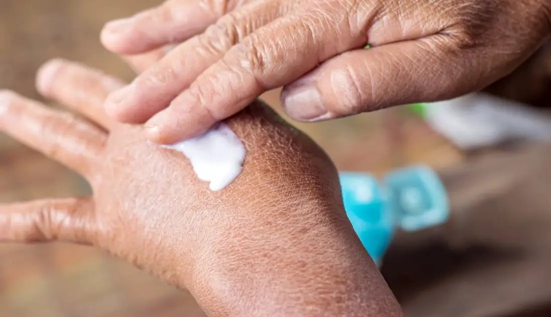
Moisturizing represents the foundation of keratosis pilaris management. Regular use of thick, occlusive moisturizers helps maintain skin hydration and can significantly improve the appearance and texture of affected areas. Moisturizers containing ceramides, hyaluronic acid, or glycerin are particularly effective.
Gentle exfoliation can help remove the keratin plugs that create the characteristic bumps. However, it’s important to avoid aggressive scrubbing, which can worsen inflammation and make the condition more noticeable. Chemical exfoliants are generally more effective and less irritating than physical scrubs.
Topical treatments containing alpha-hydroxy acids (like lactic acid or glycolic acid), beta-hydroxy acids (like salicylic acid), or urea can help dissolve keratin plugs and improve skin texture. These ingredients are available in both over-the-counter and prescription formulations.
For more stubborn cases, dermatologists may prescribe topical retinoids, which help normalize skin cell turnover and can significantly improve keratosis pilaris over time. However, these medications can be irritating initially and require careful introduction.
Laser therapy and other professional treatments may be considered for severe cases that don’t respond to topical treatments. However, these approaches are typically reserved for cases where the cosmetic impact is severe and other treatments have been unsuccessful.
Prevention Strategies: Protecting Your Skin’s Future
Sun Protection: The Foundation of Prevention
For actinic keratosis prevention, comprehensive sun protection represents the most important intervention. This protection must be consistent and lifelong, as UV damage is cumulative and irreversible.
Sunscreen application should become a daily habit, regardless of weather conditions or planned activities. Broad-spectrum sunscreens with SPF 30 or higher provide effective protection when applied properly and reapplied regularly. Many people underestimate the amount of sunscreen needed—approximately one ounce for the entire body is the recommended amount.
Physical sun protection through clothing, hats, and sunglasses provides additional protection beyond sunscreen. Tightly woven fabrics, wide-brimmed hats, and UV-blocking sunglasses all contribute to comprehensive protection. Some clothing is specifically designed with UV protection and can be particularly valuable for people at high risk.
Seeking shade during peak UV hours (typically 10 AM to 4 PM) significantly reduces exposure risk. This is particularly important for people with fair skin or those with a history of sun sensitivity.
Avoiding tanning beds entirely is crucial, as these devices expose users to concentrated UV radiation that significantly increases skin cancer and actinic keratosis risk. There is no such thing as a “safe tan” from artificial UV sources.
Genetic Counseling and Family Planning
For conditions with strong genetic components like keratosis pilaris and seborrheic keratosis, understanding family history can help guide prevention and screening strategies. While genetic testing isn’t typically necessary for these conditions, knowing your family history helps set appropriate expectations and screening schedules.
Genetic counseling may be valuable for families with multiple members affected by skin conditions or skin cancers. This can help identify inherited cancer syndromes or other genetic factors that might influence skin health.
For pregnant women with keratosis pilaris, understanding the genetic implications can help prepare for potential skin changes in children and establish appropriate care routines early.
Lifestyle Modifications
Maintaining overall skin health through proper nutrition, hydration, and gentle skin care practices benefits all types of keratosis. A diet rich in antioxidants may help protect against UV damage, while adequate hydration supports healthy skin barrier function.
Regular skin self-examinations help identify new or changing lesions early. Learning to recognize your normal skin patterns and noting any changes can facilitate early detection and treatment of concerning developments.
Establishing relationships with qualified dermatologists for regular professional skin examinations is particularly important for people at high risk for actinic keratosis or with strong family histories of skin conditions.
When Professional Help Is Essential
Red Flag Symptoms
Certain symptoms warrant immediate professional evaluation, regardless of your comfort level with self-diagnosis. Any lesion that bleeds spontaneously, grows rapidly, becomes painful, or changes significantly in color or texture should be evaluated promptly.
For actinic keratosis, lesions that become thick, inflamed, or develop open sores may indicate progression toward squamous cell carcinoma and require urgent evaluation.
New growths that appear different from your existing skin lesions should also be evaluated, particularly if they don’t fit the typical patterns of common benign conditions.
The Importance of Professional Monitoring
Even when initial treatment is successful, ongoing monitoring remains important for many types of keratosis. Actinic keratosis, in particular, requires regular follow-up to monitor for recurrence and to identify new lesions.
Professional skin examinations can identify concerning changes that might not be apparent to patients, particularly in hard-to-see areas like the scalp or back.
Dermatologists can also provide guidance on evolving treatment options and prevention strategies, helping patients adapt their care as new research emerges.
Living with Keratosis: Long-term Management and Expectations
Realistic Expectations
Understanding what to expect from keratosis treatment helps set realistic goals and reduces frustration with the management process. Many types of keratosis are chronic conditions that require ongoing management rather than one-time cures.
For seborrheic keratosis, new lesions may continue to appear over time, requiring periodic treatment. This is normal and doesn’t indicate treatment failure.
Actinic keratosis treatment success is measured not just by clearing existing lesions but by preventing new ones and avoiding progression to skin cancer. This long-term perspective is important for maintaining motivation for ongoing prevention efforts.
Keratosis pilaris improvement is often gradual and may require several months of consistent treatment to see significant results. Patience and persistence are key to successful management.
Quality of Life Considerations
For many people, keratosis affects quality of life primarily through its impact on appearance and self-confidence. Addressing these psychological aspects is an important part of comprehensive care.
Support groups and online communities can provide valuable emotional support and practical tips for managing keratosis conditions.
Working with dermatologists who understand the cosmetic impact of these conditions can help ensure that treatment plans address both medical and psychological needs.
Conclusion: Empowering Yourself Through Knowledge
Understanding keratosis in all its forms empowers you to make informed decisions about your skin health. Whether you’re dealing with benign cosmetic concerns or potentially serious precancerous changes, knowledge provides the foundation for effective management and peace of mind.
The key takeaway is that most skin changes are manageable when properly identified and treated. Early intervention, consistent prevention strategies, and regular professional monitoring can help maintain healthy skin throughout your lifetime.
Remember that your skin health is an investment in your overall well-being. The time and effort spent on proper care, protection, and monitoring pays dividends in both health outcomes and quality of life. When in doubt, professional consultation provides the expertise needed to navigate complex skin health decisions confidently.
By staying informed, maintaining good prevention habits, and seeking appropriate professional care, you can effectively manage keratosis conditions and maintain healthy, comfortable skin for years to come.

Adrian Hawthorne is a celebrated author and dedicated archivist who finds inspiration in the hidden stories of the past. Educated at Oxford, he now works at the National Archives, where preserving history fuels his evocative writing. Balancing archival precision with creative storytelling, Adrian founded the Hawthorne Institute of Literary Arts to mentor emerging writers and honor the timeless art of narrative.
Cheers!